
Positron Emission Tomography PET Scans
Positron emission tomography (PET) scans play a crucial part in the diagnosis and management of cancer. This advanced technology enables doctors to see what is happening with metabolism within the body, thus allowing them to identify and characterize tumor tissues accurately.
Working Process of Positron Emission Tomography (PET) Scans
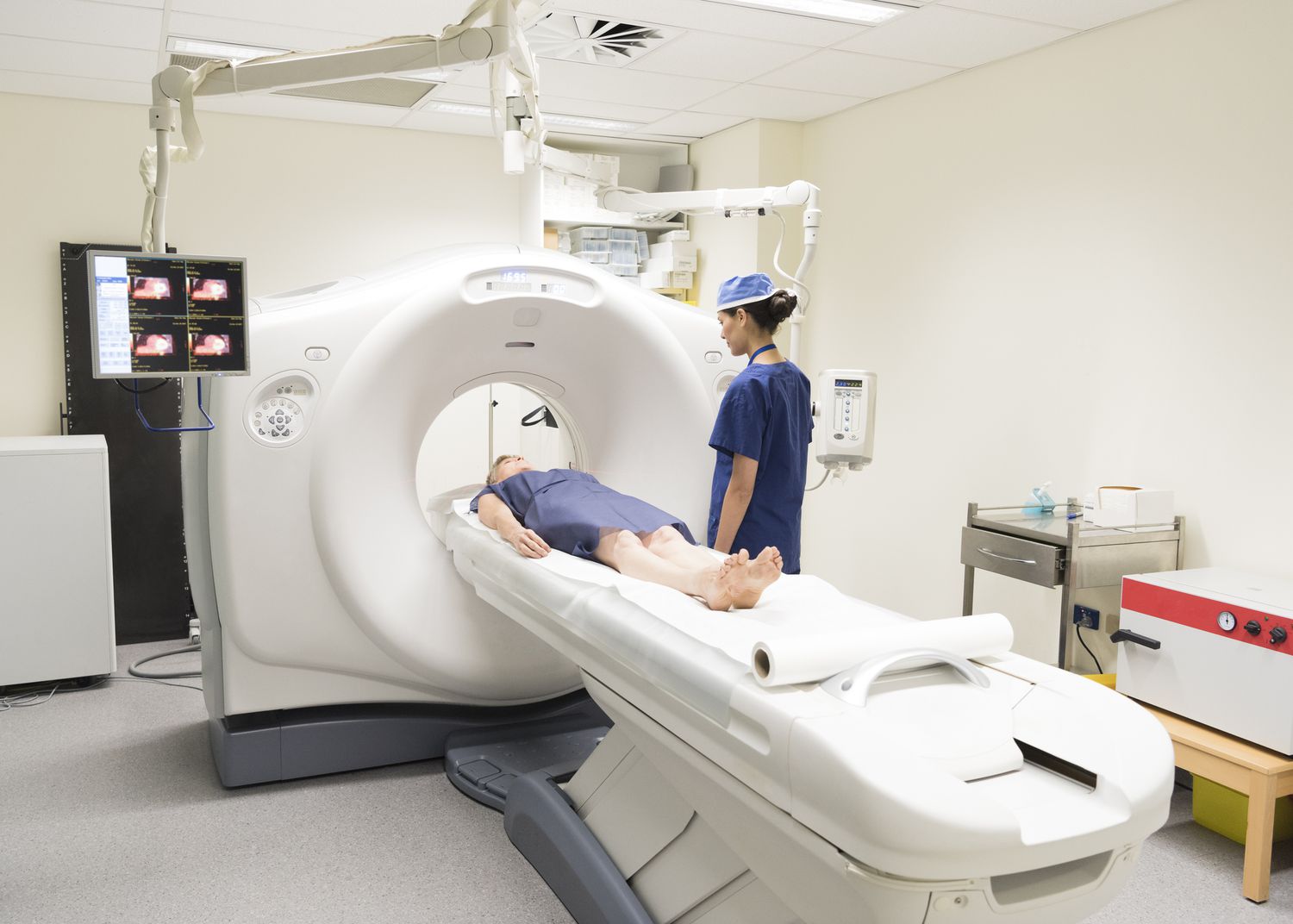
Positron Emission Tomography starts by injecting a radioactive substance into the patient’s body. Cancer cells absorb these substances more since they have higher metabolic rates than normal cells in our bodies. The tracer then needs about an hour before it can be distributed all over the body; this time, the patient is requested to rest so that they do not carry out any activities for faster distribution. Afterward, patients are taken into a PET scanner, which makes images showing how these tracers are distributed through the human body.
Specialists examine these images to search for areas of abnormal metabolic activity that may suggest cancer. PET scans are excellent at differentiating between benign and malignant tumors.
Role of PET scans
Another role PET scans play in personalized medicine is that doctors can develop individualized treatment strategies based on detailed knowledge about tumor biology for each patient when examining FDG PET results. This can go a long way toward optimizing outcomes and minimizing unnecessary treatments.
Positron Emission Tomography (PET) scans are an invaluable tool in the fight against cancer. They offer a non-invasive, detailed, and effective means of diagnosing and managing cancer, facilitating early intervention, and personalized treatment plans. By leveraging this advanced imaging technology, healthcare providers can significantly improve cancer patients’ quality of care and outcomes.
PET scans are essential diagnostic tools for oncology. They provide crucial information on detecting cancers accurately and staging them accordingly among cancer cells that display increased metabolism rates. These tracers bind to glucose-hungry areas within tumors, providing accurate images of them.
After being injected with a tracer substance through their veins, there is a waiting period during which it is evenly distributed throughout their entire body systems. The emitted positrons interact with the electrons inside the human body, leading to gamma rays detected by patients’ placement in a PET scanner. Machines capture these γ-rays and generate images illustrating where someone has cancer or its extent.
In summary, as a powerful diagnostic tool with few side effects, PET Scans should make patients aware of what happens before they undertake the process to have a great experience and result.
Frequently Asked Question
The PET scan is foremost the best imaging technique used in the diagnosis of cancer. It helps doctors visualize the tissues and organs under observation and their workings. In oncology, the most efficient advantage of this scan rather than other tests based on anatomical changes alone is that these scans show both where there may be malignancy and how quickly cancer cells consume materials such as glucose.
There are some measures which we should consider. First of all, patients have to be on fast before these PET scans as there will be no stomach upset and disturbance in the working of the tracer during the scan if food is present. It is the only preparation and prevention that needs to be considered before the examination.
In PET scans, patients are introduced to a small amount of tracer. The tracer is usually glucose, and when it enters the patient’s blood, it starts getting metabolized by the cancer and tumor cells. This process usually takes about an hour, as this is the least time required for the circulation of a radioactive substance.
PET scans are often safe without serious adverse effects. However, although radiation doses tend to be low and excreted from the body rapidly, it may still be essential to inform your doctor about your health situation or if you are pregnant as a precautionary measure. Just like when receiving injections, some people might notice coldness or mild pain that results from the presence of these substances within their veins.
After performing this scan, a radiologist will interpret the results and present a comprehensive report to your physician, revealing some trace uptakes. The report mostly shows rapid metabolic rates typical for cancer cells.
